
Jaw bone tori, or “oral tori,” are small, benign bony growths that develop inside the mouth—usually along the lower jaw or the roof of the mouth. For most people, these growths go unnoticed until they cause discomfort or become visible beneath the gum line. The searcher’s intent here is to understand what jaw bone tori are, whether they are dangerous, and how they can be treated or managed effectively. The truth is, jaw bone tori are not harmful tumors or infections; rather, they are natural bone growths that can occur due to genetic, environmental, or mechanical factors like teeth grinding or jaw pressure. They are more common than most people realize and can vary greatly in size, shape, and occurrence.
To the untrained eye, jaw bone tori may look unusual or alarming, but they are generally harmless. Some individuals discover them during routine dental checkups, while others notice them when brushing or eating. While not life-threatening, tori can complicate oral hygiene, denture fitting, and even speech in some cases. Understanding what triggers these bony growths and when to seek professional help can empower individuals to manage their oral health proactively. In recent years, dentistry has developed new insights into their causes and treatment, shifting away from unnecessary surgical interventions toward conservative monitoring and lifestyle adjustments. “The mouth is a mirror of the body’s balance,” says Dr. Ellen Proctor, a maxillofacial specialist. “Tori often reflect how our bones adapt to stress, heredity, and oral habits.”
What Are Jaw Bone Tori?
Jaw bone tori are noncancerous bone outgrowths covered by thin mucosal tissue. These bony protrusions are classified into two main types: tori mandibularis, found on the inside of the lower jaw near the tongue, and tori palatinus, located along the midline of the hard palate. Occasionally, less common variants such as buccal exostoses appear along the outer gum line of the upper or lower jaws. These growths consist primarily of dense cortical bone and can remain stable for years or slowly enlarge over time. Their texture is typically hard to the touch and smooth under the gum layer. The prevalence of tori varies by ethnicity and genetic background, with higher rates observed in certain populations, particularly those of Asian, Inuit, and Scandinavian descent.
Table 1: Common Locations and Characteristics of Jaw Bone Tori
| Type of Tori | Location | Texture | Typical Size | Occurrence Rate | Symptoms |
|---|---|---|---|---|---|
| Tori Mandibularis | Inside lower jaw, near tongue | Hard, smooth | 2–5 mm (can enlarge) | 7–10% of population | Rarely painful |
| Tori Palatinus | Roof of mouth (midline) | Dome-shaped, hard | 3–8 mm average | 20–30% of population | Can irritate with dentures |
| Buccal Exostoses | Outer gum line (upper/lower) | Irregular surface | 1–4 mm | Less than 5% | May affect cheek lining |
Jaw bone tori generally begin forming in early adulthood and may remain unnoticed for decades. They grow slowly and rarely cause any serious health problems, but they can interfere with prosthetic dental devices, particularly dentures or aligners. They are not a result of infection or trauma; instead, they emerge as part of natural bone development or response to physical stress, much like calluses form on the skin.
Causes of Jaw Bone Tori
The exact cause of jaw bone tori remains multifactorial. Genetics play a major role—individuals with a family history of tori are significantly more likely to develop them. Additionally, mechanical stress from teeth grinding (bruxism), clenching, or heavy chewing may stimulate localized bone growth. Hormonal influences, dietary calcium intake, and overall bone density are also contributing factors. Some studies suggest that repeated microtrauma from biting or poor dental alignment may encourage the bone to thicken over time.
Environmental conditions and lifestyle habits also influence tori development. For instance, populations with diets high in fibrous or tough foods tend to show higher occurrences of these growths due to continuous jaw stimulation. “Our bones adapt to the stress they experience,” explains Dr. Nathanial Rhodes, a dental anthropologist. “Jaw bone tori are a fascinating example of how daily habits shape our anatomy over decades.” Interestingly, there is no evidence linking tori to oral cancer or infections; rather, they are simply exaggerated bone responses to normal mechanical loads.
Recognizing the Symptoms
Most people with jaw bone tori experience no pain or symptoms. However, larger tori can occasionally cause irritation, ulceration, or difficulty with oral appliances. They may also interfere with speaking, eating, or cleaning if food debris becomes trapped around them. Some patients report tenderness if the mucosal covering becomes thin or injured, often from sharp food particles or ill-fitting dentures. In rare cases, tori can become secondarily infected when small cuts in the overlying tissue expose the underlying bone.
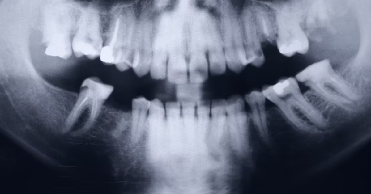
The early signs usually appear as firm bumps or ridges beneath the gum, particularly on both sides of the lower jaw. Tori can be unilateral (one-sided) or bilateral (both sides). They tend to grow very slowly and may remain the same size for many years. Dentists can easily identify tori through visual inspection and X-ray imaging, ensuring that they are not mistaken for cysts, tumors, or other pathologies.
When to Seek Dental Attention
Although jaw bone tori are benign, professional evaluation is crucial when they cause discomfort or interfere with dental work. Dentists typically monitor the size and shape of tori over time to ensure they remain stable. Surgery is rarely recommended unless the growths obstruct speech, swallowing, or prosthetic fitting. For patients requiring dentures or implants, tori removal may be necessary to achieve a smooth surface for attachment.
Patients are advised to maintain proper oral hygiene, as plaque accumulation around the tori can irritate the surrounding tissue. Regular dental visits ensure that any changes are detected early. It is also essential to avoid self-diagnosis; while tori are harmless, other conditions like osteomas or cystic lesions may present similarly. “A simple panoramic X-ray can confirm the diagnosis,” notes Dr. Lydia Kim, oral radiologist. “Understanding whether a growth is physiological or pathological can prevent unnecessary anxiety.”
Treatment Options and Recovery
Treatment for jaw bone tori depends on the severity and symptoms. In most cases, no intervention is needed. However, when surgical removal becomes necessary, it involves a minor outpatient procedure known as torus reduction. This process includes lifting the gum flap, reshaping or trimming the bone, and suturing the tissue. Recovery typically takes two to four weeks, depending on the patient’s health and oral hygiene. Post-operative care includes soft foods, antiseptic mouth rinses, and minimal physical strain on the surgical area.
The advancements in laser-assisted and minimally invasive bone contouring techniques have made the procedure more comfortable and precise. Dentists today use digital mapping to plan the surgery, ensuring minimal tissue trauma. The recurrence of tori after removal is rare but possible, particularly if the underlying cause—like bruxism—remains unaddressed. Preventive dental guards, jaw relaxation exercises, and stress management techniques can help mitigate these risks.
Table 2: Treatment and Management Approaches
| Condition Severity | Recommended Action | Procedure Type | Recovery Time | Recurrence Risk |
|---|---|---|---|---|
| Mild (asymptomatic) | Observation, regular dental check | None | None | Low |
| Moderate (irritation) | Adjust dentures, improve hygiene | Non-surgical | 1 week | Low |
| Severe (pain or obstruction) | Surgical removal (torus reduction) | Outpatient procedure | 2–4 weeks | Rare |
| Post-surgery maintenance | Mouth rinses, soft diet, guard use | Supportive care | 2 weeks | Very low |
Preventive Strategies and Home Care
While you cannot completely prevent jaw bone tori, certain lifestyle choices can minimize their progression. Reducing teeth grinding through mouthguards, managing stress, and avoiding chronic jaw clenching are effective measures. Maintaining balanced calcium intake supports healthy bone metabolism without overstimulation. People with tori should also be cautious when eating hard or abrasive foods to prevent gum injury. Regular professional cleanings and dental assessments play a significant role in managing oral balance.
Those who wear dentures should ensure proper fitting, as friction against the tori can lead to inflammation. Using soft toothbrushes and avoiding harsh brushing near bony areas can also protect the thin mucosa. Moreover, a dentist can customize protective guards for individuals prone to bruxism, thereby reducing mechanical load on the bone. As one dental maxim wisely states, “Prevention begins not in the mirror, but in mindful daily habits.”
Psychological Impact and Patient Perception
Although jaw bone tori are benign, they can create anxiety due to their unfamiliar appearance. Many patients initially mistake them for tumors or cancerous lesions, prompting emotional distress. Education plays a vital role in reducing such fears. When dentists take time to explain the condition clearly, patient satisfaction and trust significantly increase. “The moment I understood it wasn’t dangerous, my fear disappeared,” recalls one patient who discovered bilateral mandibular tori during a cleaning. This emotional reassurance underlines the importance of patient-centered dental care, where empathy accompanies expertise.
Advances in Diagnosis and Imaging
Modern imaging technologies have revolutionized how oral tori are identified and monitored. Cone beam computed tomography (CBCT) offers precise 3D visualization, allowing dentists to evaluate the exact volume and location of bone growth. Digital scanners now help create accurate oral maps for prosthetic adjustments without invasive impressions. These innovations contribute to early detection, precise diagnosis, and safer interventions when required. The future may also bring bio-molecular analysis that can reveal how genetic markers influence torus development—linking oral anatomy to personalized medicine.
Long-Term Outlook and Lifestyle Adaptation
Most people with jaw bone tori lead perfectly normal lives. The growths rarely interfere with eating or speaking, and with proper dental supervision, complications remain minimal. Those with removable prosthetics can collaborate with dental technicians to design devices that accommodate the bony contours comfortably. A long-term strategy involves consistent hygiene, stress control, and nutritional balance. As Dr. James Holloway aptly summarizes, “Oral tori are reminders of the body’s resilience—they grow quietly, adapt silently, and rarely demand attention unless we neglect them.”
Conclusion
Jaw bone tori, while often misunderstood, are among the most intriguing examples of how the human body adapts to daily mechanical and genetic influences. They represent benign yet biologically meaningful changes that reveal much about our evolutionary and lifestyle patterns. Understanding them fosters not only better oral health but also deeper appreciation for the delicate equilibrium of the mouth’s anatomy. For patients, awareness eliminates fear; for dentists, it encourages precision and compassion. As dental science progresses, the focus shifts toward maintaining harmony rather than intervention—preserving nature’s quiet architecture within our bones. Ultimately, managing jaw bone tori is less about correction and more about understanding the subtle dialogue between habit, heredity, and health.
FAQs
1. Are jaw bone tori dangerous or cancerous?
No, jaw bone tori are completely benign and noncancerous. They are natural bone growths that do not transform into malignant tumors or spread to other areas.
2. Can jaw bone tori disappear on their own?
Tori do not typically regress naturally, but they may stop growing once bone development stabilizes. Surgical removal is the only way to eliminate them completely.
3. Is surgery for jaw bone tori painful?
The procedure is generally painless under local anesthesia. Post-operative discomfort is mild and manageable with prescribed pain relief.
4. Can dentures be worn over tori?
Yes, but adjustments may be needed for comfort and fit. In some cases, surgical reduction is advised before denture placement.
5. How common are jaw bone tori?
They are more common than most people think—occurring in about 10–30% of adults globally, with variations across populations.